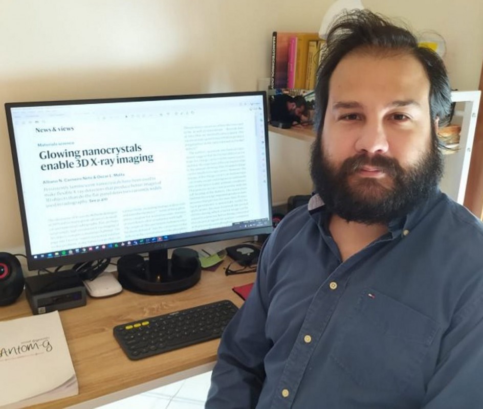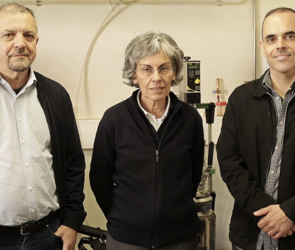
It is a new technology that wants to revolutionize the images obtained through radiography using nanocrystals containing lanthanide ions that produce a luminescence prolonged in time. In an article named "Glowing nanocrystals enable 3D X-ray imaging" published in Nature journal, the researcher Albano Carneiro from CICECO-Aveiro Institute of Materials and Department of Physics from University of Aveiro (UA), in collaboration with the researcher Oscar Malta, from the Federal University of Pernambuco (Brazil), reports the advances that this new technology born in Singapore can bring to the field of medicine and materials.
Since the discovery of X-rays by Wilhelm Conrad Röntgen (winner of the 1901 Nobel Prize in Physics), advances have been made in the medical and industrial fields. Today, even the most advanced detection techniques in radiography still use non-flexible flat plates that convert the absorbed energy (from X-ray radiation) into electronic signals. “The resolution of the radiographic image can be limited because of the use of these flat plates that, in some cases, can reveal overlaps between faces of the same object,” explains Albano Carneiro.
To circumvent this three-dimensional (3D) resolution gap, a new technology called X-ray luminescence extension imaging (Xr-LEI) has been developed by the team of Xiaogang Liu, a scientist at the National University of Singapore. This technology consists of a new proposal to replace conventional detector plates with a flexible silicon-based material in which nanocrystals containing lanthanide ions are incorporated.
These nanocrystals, explains the scientist from UA, “when hit by X-rays, produce structural defects at the atomic level (small displacements in the position of some atoms), which are responsible for storing the energy absorbed by the material.” After the end of the irradiation, “these atoms slowly return to their original position transferring part of the absorbed energy to the lanthanide ions that, in turn, emit light that can persist for 30 days.”
“Because it is based on a flexible material, the Xr-LEI detector can be shaped to fit the 3D object to be evaluated. In addition, better resolution images are obtained, relative to those obtained with conventional detectors, with the advantage of requiring a shorter X-ray exposure time (around 1 second). There are many perspectives for this technology in the area of medical diagnosis, as well as in the inspection of electronic circuits,” predicts Albano Carneiro.
Related Articles
We use cookies for marketing activities and to offer you a better experience. By clicking “Accept Cookies” you agree with our cookie policy. Read about how we use cookies by clicking "Privacy and Cookie Policy".


